Friday, March 27, 2015

Week 10. Semester 2. Failed.

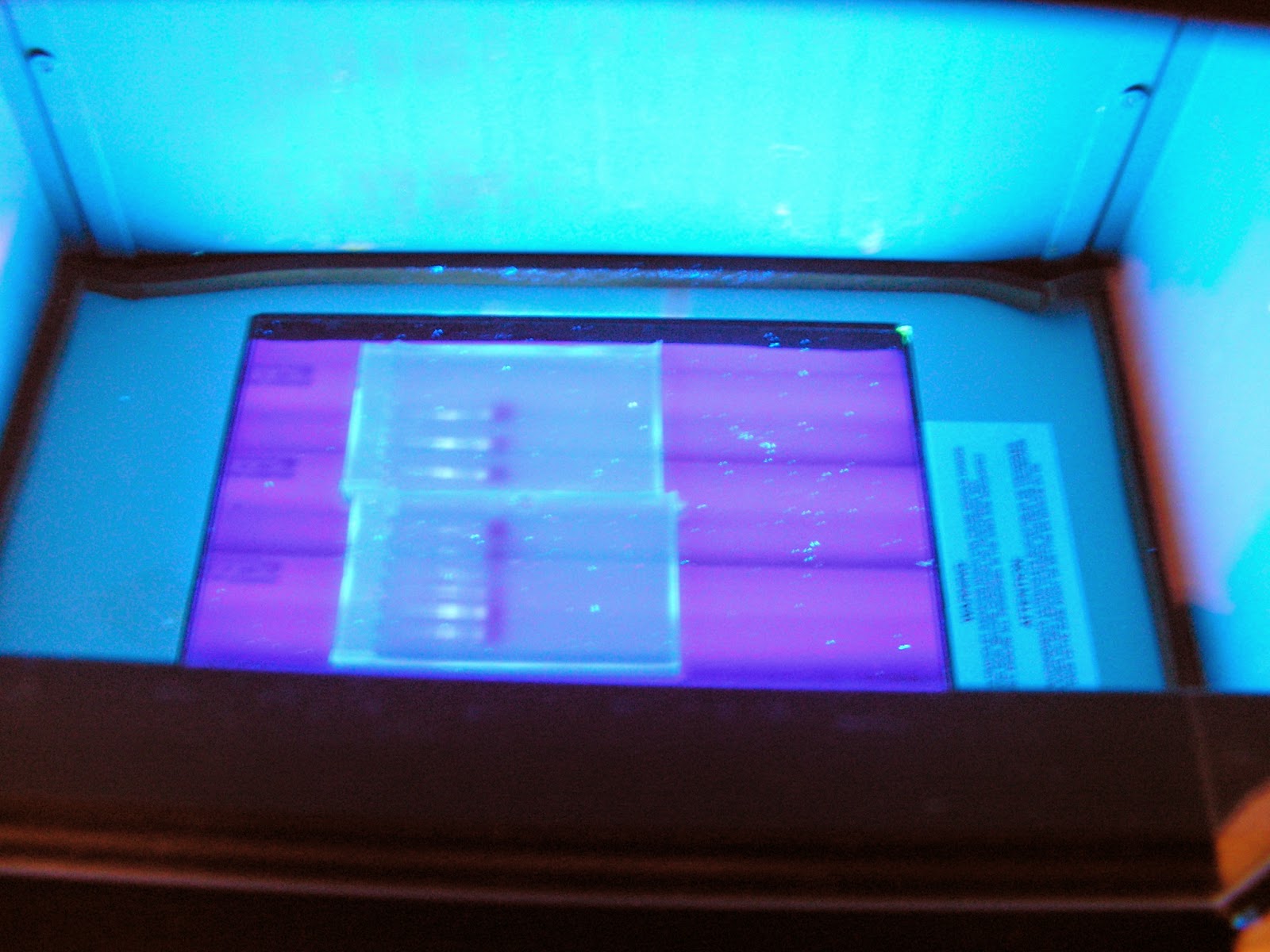
After incorporating an incubation period of 1 hr and overnight, respectively, in a solution containing Aspergillus cellulase complex to my previously used protocol, no DNA could be detected under UV light.

Unfortunately, there were a few mishaps in this run. To list a few:

- Hot plate was left on at 65 degrees Celsius for the overnight incubation, but I failed to leave a note and after Mat (the only one aware) left, someone turned it off, which probably was the right thing to do since it had no "please don't turn off" note.

- After the cellulase and lysis buffer incubations, there is a centrifuge step and the supernatant is to be kept because all the cellular debris pellets in the bottom. Accidentally I discarded the supernatant.

- For the overnight extraction, there are two PCI then chloroform extractions both followed by an alcohol addition step. To save time I thought I would synchronize the extractions and ended up unintentionally skipping the second PCI & chloroform extraction.

-The Optimal pH (5.0) and Temp (65C) I used for the cellulase incubation were based on information provided from the Manen et all. 2005 research article I cited on my last post. The source organism was the fungus Trichoderma. While recording my results I found a note I had made to myself about the optimal pH and Temp of Aspergillus. Definitely not the same...


All things considered I kinda knew I wouldn't find anything. For next week I will modify the incubation time and temperature, as well as the cellulase buffer and see what happens. Please take a look at my failed extractions. The one showing DNA is actually my control from a previous extraction.
   Only bottom has visible DNA and is from previous extraction.

I chose to have a Grind and No Grind sample.

Tuesday, March 17, 2015

Week 8. Semester 2. Will Palo Verde cell walls hold?


Last week I spent a lot of time doing research on the celullase complex and the structure of cellulose itself. Mat and I are still trying to come up with a way to determine whether the cell wall has been degraded and to what extent by the cellulase solution before starting the extractions. Initially, we thought we may use the Maltose color reagent and spectrophotometry to generate a curve to find the optimal pH and Temperature within a range that won't denature DNA molecules. So far we have the optimal pH of the complex at between 4-5 and temp at 60-70 C, but the low pH may cause severe damage to DNA. So far I think we are going to keep the pH at 5 for the digestion time and then proceed with the buffer and the same protocol I have been using. I will be carrying out two extractions, one with chopped pieces of tissue and the other one with ground with mortar and pestle as usual.
These couple of days we get free I have been wondering whether this could work. Ideally it will be a very nice addition to the protocol, that is to have enzymes aid in the breakage of cellulose. However, as of today I have not found any information on any fungus attacking Palo Verde trees, except for one that grows in its roots. So all my planing may actually not even work. Either way, I have become very interested on enzymes and specially those in fungi. So although this may or may not work for this project, it may be the basis of my own project next semester.

 The though Sonoran desert tree may not even be susceptible to enzymatic digestion
 
This is so funny... I must add it. As I was looking for the info from the picture to cite, I realized it was pulled from our own Matthew H's blog: Practical Biology: Science for Everyone. Available at: http://practicalbio.blogspot.com/

Thursday, March 12, 2015

Week 7. Semester 2. Research.

This week was spent doing research. I have concluded, with Matt's assistance and support that we will focus on isolating cellulases from fungi instead of anaerobic bacteria due to the difficulties we may face attempting to first, successfully grow anaerobic bacteria in the lab. And secondly, to extract cellulase complexes from bacteria. Because fungus secrete enzymes into their environment, they are a better candidate for this project because the exoenzymes would be present in the medium and could be isolated using the centrifuge and molecular filters (Manen et all. 2005). I am following Bethany's, a fellow S-STEM scholar, project closely because she is growing fungus for isolation and identification. Once she is able to find either Aspergillus or Trichoderma, the broth will be started.
The lab also purchased cellulase from Sigma in solution and a powder from from another supplier, so I also spent this week learning about cellulase and how it can be incorporated in my protocol. Hopefully after Spring break I will attempt an extraction using cellulase to digest the cell wall. There is still much research and experimentation I must do in order to carry out an effective extraction. Cellulase is the main component of plant cell walls and is a very rigid polysaccharide that maintains the cell shape of plant cells and provides additional structural support. As you may have read in my previous blogs, this is one of the main constraints to extracting high yields of DNA from plant material (Manen et all. 2005). Below are some pictures of cellulose that would hopefully aid to better describe its composition and toughness.

Colored transmission electron micrograph of cells in a young leaf (Robinson 2001).


Structure of Cellulose in a Plant Cell Wall (Moran-Mirabal 2003)

References
     
      Manen JF, Sinitsyna O, Aeschbach L, Markov AV, Sinitsyn A. 2005. A fully automatable enzymatic method for DNA extraction from plant tissues. BMC Plant Biology [Internet]; [cited Mar 12 2015]. Nov 3;5;23. Available from http://www.ncbi.nlm.nih.gov/pubmed/16269076.  Manen JF, Sinitsyna O, Aeschbach L, Markov AV, Sinitsyn A.
      Moran-Mirabal J. 2003. Advanced-microscopy techniques for the characterization of cellulose structure and cellulose-cellulase interactions. In: Cellulose - fundamental aspects [Internet]; [cited Mar 12 2015]. Available from: http://www.intechopen.com/books/cellulose-fundamental-aspects/advanced-microscopy-techniques-for-the-characterization-of-cellulose-structure-and-cellulose-cellula  

      Cosgrove DJ. 2001. Cell walls. In: Plant Sciences [Internet]; New York: Macmillan Reference USA; [cited Mar 12 2015]. Available from: http://ezproxy.pc.maricopa.edu:2048/login?url=http://go.galegroup.com/ps/i.do?id=GALE%7CCX3408000066&v=2.1&u=mcc_phoe&it=r&p=GVRL&sw=w&asid=75046d4a30a1cb609c50d446a88f427f


Thursday, March 5, 2015

Week 6. Semester 2. Another extraction.

This week another DNA extraction was performed. This time I used tubes pre-loaded with Phase Lock, a Vaseline like gel that has a density in between the DNA solution and phenol-chloroform, which helps keep the phases separated. I personally do not believe it was as helpful as I had anticipated but I will try using it once more, next time centrifuging the tubes before using them. Shipping may have contributed to the gel getting spread out in the tube and I think that hindered more than helped my extraction. I also found a possible DIY protocol to make a gel similar to Phase Lock, I may try that as well next week.
Instead of extracting DNA from one of the practice samples in the lab, this week I decided to extract DNA from a Palo Verde tree behind the west corner of the library on campus. I believe having the fresh specimen collected and proceeding to extract DNA immediately may contribute to higher amounts of isolated DNA. Please refer to the images below for the results of the extraction.


Both from same specimen and protocol. Difference may be due to difference in
concentrations of DNA solution due to the fact I drop one of my tubes at some point
in the extraction and lost a bit of aqueous solution.


Lines 1 & 2 are from last extraction, from tree on SW corner of library.
Lines 3-5 are from practice specimen labeled IIIC.
Lines 6-8 are from practice specimen IIIA.


Wednesday, February 25, 2015

Week 5. Semester 2. DNA extraction success!!

Last week I was unable to run another practice extraction due to the holiday. This week however, I was able to complete my extraction in one day! If you recall from my first time doing one, it took me a whole week to complete it. Today I electrophoresed a couple of gels, please refer to images below. I have found adding short centrifugations in the protocol after addition of ethanol for precipitation helps to more effectively isolate the DNA. When loading the gels I used orange loading dye instead of the usual green/blue dye and found it to interfere less with the luminescence of cyber green under UV light. Please enjoy the pictures below! I am very excited about my results and although not compared to seeing it live, I hope you share some of the excitement!
For next week, my plan of action will depend on the arrival of cellulase and PCR primer. I am curious to try alternative extractions. But also to move ahead in the project and test the extractions yield and quality for PCRs.


This week's DNA extraction from practice sample IIIC 


Last extraction from practice sample IIIA shown in first upper lines
this week's extraction from IIIC shown in three lower lines


Two gels in UV viewer
As close as you can get to seeing it live


Tuesday, February 17, 2015

Week 4. Semester 2. Some DNA!!

As you may recall from last week, my extraction was finished but I was unable to determine whether I had DNA before submitting my post.

The following was done Friday morning last week:

Upon retrieving the DNA samples suspended in TE buffer from the refrigerator, two agar gels were prepared with 50 mL of 1x TAE buffer and .450 g of agarose. Once cooled to 59◦C, 4µL of Cyber Green were added to the beaker immediately before pouring into gel molds. A cardboard box was used to cover the gels and protect the Cyber Green because it decays when exposed to light and heat.

Once the gels were set, 2µL of Loading Dye were used in each well in the gels and 4µL of DNA sample loaded accordingly. 1x TAE buffer was added to cover the gels. The gels were electrophoresed for only 10 min, due to time constraints, and very little DNA was visible. Then Matt Haberkorn kindly offered to run them again and did so for approximately another 25 min. DNA run further and very faint banding was present.

However little, having some DNA in my first independent extraction feels like a great success!!


The remainder of the week I will be researching for possible ways to add cellulase to my protocol in hopes of increasing the yield of DNA per sample.


 Faint but visible DNA banding from practice extraction

Thursday, February 12, 2015

Week 3. Semester 2. First Plant Extraction. Have something?

This week I was able to perform an extraction using plant tissue Matt had saved for practice runs. During the extraction I encountered quite a few conflicts with the information in the protocol I had and Matt's protocol. Eventually I found out he had most of his adjustments in the protocol described in his research paper. So lesson learned, I double checked in the paper before moving ahead with the next step in my protocol.
This morning I finished the extraction and although it looked as if I had nothing (a DNA pellet was supposed to be present in the alcohol solution). I centrifuged it for 5 min. and voula! There was a visible precipitate. I am not sure it is DNA. I actually think it's not because of it's color. However, I will run an electrophoresis tomorrow to check.
So I know I have something, I'll find out tomorrow whether it is DNA.
I will be discussing my results next week.

Improvised water bath for lysis buffer and set up to grind plant tissue sample


First time working with Phenol and Chloroform


Barely visible precipitate on dried tube (hoping this is DNA)


DNA suspended in 1xTE buffer and stored in the refrigerator to dissolve