Wednesday, November 18, 2015

Week 11. Semester 3. PCR on samples from April 2015

PCR was performed on samples collected 04/24/2015 and extracted 04/27/2015.
The following is the protocol utilized.

Polymerase Chain Reaction (11-18-15)
PCR Tube: 20 μL MasterMix, 4 μL Forward/Reverse Primers,
2μL DNA*and 10 μL Sterile Water (for positive controls)
12μL DNA and no water (for samples)
ThermoCycler at an annealing temperature of 55 ͦC for 34 cycles with hold at  4 ͦC
Primers Used:  16s LEG-226: AAGATTAGCCTGCGTCCGAT (654 bp);
16s LEG-858: GTCAACTTATCGCGTTTGCT.
mip LpneuF: CCGATGCCACATCATTAGC (150 bp);
mip LpneuR: CCAATTGAGCGCCACTCATAG.

Samples collected 04/24/2015 coded 1-10 16s.
No optional step during extraction.
Extracted 04/27/2015
Sample No.
Location

1
Middle Football Irrigation Control Box (Biofilm)
2
South Gym Family Shower Drain (Biofilm)
3
Hacienda AC Roof Top (Biofilm)
4
DB-123 Station Z Eyewash Drain (Biofilm)
5
Center Field Football Sprinkler Head (Biofilm)
6
Football Snack Bar Fountain Head (Biofilm)
7
Hacienda Non-Potable (Biofilm)
8
North Valve Rooftop cooling Tower Sitting Water (Water)
9
F-Building East Spigot (Water)
10
Hacienda Non-Potable (Water)


Experimental Errors:

Used Master Mix from different tubes. 

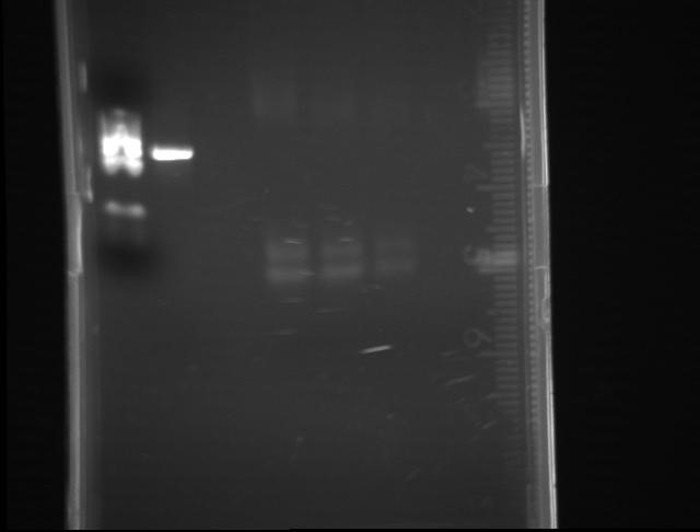
PCR amplification. An illustration showing how DNA is amplified in the first four cycles. 
Courtesy of: West Coast Pathology Lab.

Thursday, November 12, 2015

Week 10. Semester 3. Preparation for Poster Presentation

This week, Friday October 13th, the team will meet to discuss our results and how to interpret them and present them in our report for WAESO and to Dr. Schwake for review to incorporate to his research.
According to my understanding, the team will be briefed on running a BLAST on the sequences we obtained from Core Labs at ASU's School of Life Sciences. Ideally we hope to find our samples match those in the database for the microphage ineffectivity potentiator and 16s genes for Legionella. 
Additionally, this week the team will join the S-STEM Scholars from PC in a field trip to ASU West campus. We will be touring the Labs and meeting the faculty and learning about their research.

ASU West Campus
Photo courtesy of: artscare.org Available at:http://www.artscare.org/images/cac.event.46.02.jpg

Week 9. Semester 3. Preparation for Sequencing

On October 06th the DNA samples extracted from the agarose gels were taken to ASU for sequencing in order to compare the nucleotide sequence of the samples with the databases available online. The protocol for the preparation of samples follows:

DNA Sequencing (11-06-2015)
Utilizing a NanoDrop to quantify the mass of DNA in each tube, the mass for each sample was recorded in ng/mL.
First the NanoDrop was blanked with 1µL of sterile water. Only one blank was necessary for all the samples.
The apparatus was wiped with a KimWipe in between the blank and each sample.
1µL of each sample and positive control (purified from agarose gels) was used per sample.
Once the mass was recorded, the tubes were prepared as follows for sequencing:

Each sample had to contain at least 20ng per tube. The smallest mass recorded was ≈5ng/mL. Therefore, 4µL of each sample and controls were added to each labeled tube along with another volume of forward primer. Depending on the sample type, mip or 16s primer was added. 

For reference, I have attached a picture of a similar sequencing machine to the one the team got briefed on last Friday.

Photo courtesy of: ASU Biodesign Institute. Available from: http://cpdlab.biodesign.asu.edu/images/3730.jpg

Week 8. Semester 3. DNA extraction from gels


On Friday, October 27th and 28th, the team worked on extracting DNA from agarose gels coming from PCR amplified field samples. The same samples that had tested positive in the Summer banded at the mip and 16s molecular weights once more. Please refer to the pictures and tables for more info.
Agarose gel DNA Extraction for sequencing (10-17-15)

Utilized UV lamp to visualize bands. Cut with scalpel and placed in DNAase free

tubes.

Extracted DNA from gel as per the Thermo Scientific GeneJET Extraction kit:

Added binding buffer on 1mg (agarose):1μL ratio.

Incubated at 55◦C for ≈12 min (until get dissolved). Inverting at 2 min intervals.

Vortexed for 5-10 seconds.

For mip DNA only, a 1 gel volume of 100% isopropanol was added.

Transferred solution to purification column and centrifuged for 1 min at 13.2 rpm.

Flow through was discarded.

Added 100µL of Binding Buffer to column and centrifuged for 1 min at 13.2 rpm.

Discarded flow through. And centrifuged column an additional minute.

Transferred column to DNA free centrifuge tube and added 50µL of Elution Buffer.

Centrifuged for 1 minute at 13.2 rpm. Column was discarded and DNA stored at -

20°C.

mip gel
Lane 1
Lane 2
Lane 3
Lane 4
Lane 5
Lane 6
Lane 7
Lane 8
MW
mip +
empty
12w
13w
14w
15w
16w


mip and 16s gel
Lane 1
Lane 2
Lane 3
Lane 4
Lane 5
Lane 6
Lane 7
Lane 8
MW
m+
empty
9b mip
17w mip
9b 16s
17w 16s
16s +

16s gel

Lane 1
Lane 2
Lane 3
Lane 4
Lane 5
Lane 6
Lane 7
Lane 8
MW
16s +
empty
12w
13w
14w
15w
16w