Friday, December 11, 2015

Week 14. Semester 3. Wrapping up.

Today, Friday December 11th, the S-STEM Scholars will be presenting their work on campus. At the moment I am finalizing the power point presentation. 
Typically, I abstain myself from deviating from data and procedure updates in this blog. However, this week is our last in the semester and I wanted to take the time to thank the NSF and the Phoenix College Biosciences Department, especially Dr. Amanda Chapman and Dr. Rosati for making this experience possible.
Thanks to all my fellow S-STEM scholars and our wonderful mentors for their continued support in our academic and personal life.
Special thanks to Dr. Robin Cotter for her amazing passion for teaching and empowering the students in the project. To Cori for always being there for us and for all the helpful insights and techniques taught during the semester. Thanks Josh for always listening and encouraging the scholars and Matt for being so helpful in the lab. Thanks to Kim and Ana for their niceness!!
And finally, thanks to each and every one of the interns for the wonderful conversations and the support we give each other!

I am glad to plan to return next semester and continue being a part of this wonderful group of people!
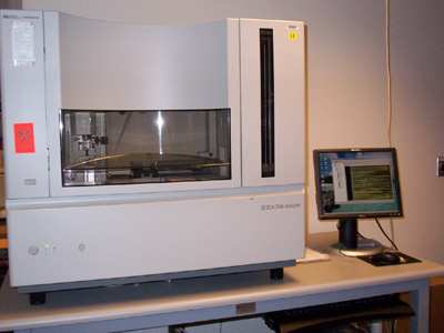
Photo courtesy of Phoenixcollege.edu

Thursday, December 3, 2015

Week 13. Semester 3. Sequencing trip to ASU

Thursday 12/03 we visited ASU to drop off samples for sequencing. The samples dropped were labeled mip 1 through 10. 

Utilizing a NanoDrop to quantify the mass of DNA in each tube, the mass for each sample was recorded in ng/mL.
First the NanoDrop was blanked with 1µL of sterile water. Only one blank was necessary for all the samples.
The apparatus was wiped with a KimWipe in between the blank and each sample.
1µL of each sample and positive control (purified from agarose gels) was used per sample.
Once the mass was recorded, the tubes were prepared as follows for sequencing:
Each sample had to contain at least 20ng per tube. The smallest mass recorded was ≈5ng/mL. Therefore, 4µL of each sample and controls were added to each labeled tube along with another volume of forward primer. Depending on the sample type, mip or 16s, 2µL primer was added.

Visiting ASU has been a very nice experience. The man running the lab, Scott is always very nice and I feel our short talks and this explanations of how sequencing works have been a meaningful part of my experience this semester.
ASU main
courtesy of finestweddingplaces.com

Wednesday, November 18, 2015

Week 11. Semester 3. PCR on samples from April 2015

PCR was performed on samples collected 04/24/2015 and extracted 04/27/2015.
The following is the protocol utilized.

Polymerase Chain Reaction (11-18-15)
PCR Tube: 20 μL MasterMix, 4 μL Forward/Reverse Primers,
2μL DNA*and 10 μL Sterile Water (for positive controls)
12μL DNA and no water (for samples)
ThermoCycler at an annealing temperature of 55 ͦC for 34 cycles with hold at  4 ͦC
Primers Used:  16s LEG-226: AAGATTAGCCTGCGTCCGAT (654 bp);
16s LEG-858: GTCAACTTATCGCGTTTGCT.
mip LpneuF: CCGATGCCACATCATTAGC (150 bp);
mip LpneuR: CCAATTGAGCGCCACTCATAG.

Samples collected 04/24/2015 coded 1-10 16s.
No optional step during extraction.
Extracted 04/27/2015
Sample No.
Location

1
Middle Football Irrigation Control Box (Biofilm)
2
South Gym Family Shower Drain (Biofilm)
3
Hacienda AC Roof Top (Biofilm)
4
DB-123 Station Z Eyewash Drain (Biofilm)
5
Center Field Football Sprinkler Head (Biofilm)
6
Football Snack Bar Fountain Head (Biofilm)
7
Hacienda Non-Potable (Biofilm)
8
North Valve Rooftop cooling Tower Sitting Water (Water)
9
F-Building East Spigot (Water)
10
Hacienda Non-Potable (Water)


Experimental Errors:

Used Master Mix from different tubes. 

PCR amplification. An illustration showing how DNA is amplified in the first four cycles. 
Courtesy of: West Coast Pathology Lab.

Thursday, November 12, 2015

Week 10. Semester 3. Preparation for Poster Presentation

This week, Friday October 13th, the team will meet to discuss our results and how to interpret them and present them in our report for WAESO and to Dr. Schwake for review to incorporate to his research.
According to my understanding, the team will be briefed on running a BLAST on the sequences we obtained from Core Labs at ASU's School of Life Sciences. Ideally we hope to find our samples match those in the database for the microphage ineffectivity potentiator and 16s genes for Legionella. 
Additionally, this week the team will join the S-STEM Scholars from PC in a field trip to ASU West campus. We will be touring the Labs and meeting the faculty and learning about their research.

ASU West Campus
Photo courtesy of: artscare.org Available at:http://www.artscare.org/images/cac.event.46.02.jpg

Week 9. Semester 3. Preparation for Sequencing

On October 06th the DNA samples extracted from the agarose gels were taken to ASU for sequencing in order to compare the nucleotide sequence of the samples with the databases available online. The protocol for the preparation of samples follows:

DNA Sequencing (11-06-2015)
Utilizing a NanoDrop to quantify the mass of DNA in each tube, the mass for each sample was recorded in ng/mL.
First the NanoDrop was blanked with 1µL of sterile water. Only one blank was necessary for all the samples.
The apparatus was wiped with a KimWipe in between the blank and each sample.
1µL of each sample and positive control (purified from agarose gels) was used per sample.
Once the mass was recorded, the tubes were prepared as follows for sequencing:

Each sample had to contain at least 20ng per tube. The smallest mass recorded was ≈5ng/mL. Therefore, 4µL of each sample and controls were added to each labeled tube along with another volume of forward primer. Depending on the sample type, mip or 16s primer was added. 

For reference, I have attached a picture of a similar sequencing machine to the one the team got briefed on last Friday.

Photo courtesy of: ASU Biodesign Institute. Available from: http://cpdlab.biodesign.asu.edu/images/3730.jpg

Week 8. Semester 3. DNA extraction from gels


On Friday, October 27th and 28th, the team worked on extracting DNA from agarose gels coming from PCR amplified field samples. The same samples that had tested positive in the Summer banded at the mip and 16s molecular weights once more. Please refer to the pictures and tables for more info.
Agarose gel DNA Extraction for sequencing (10-17-15)

Utilized UV lamp to visualize bands. Cut with scalpel and placed in DNAase free

tubes.

Extracted DNA from gel as per the Thermo Scientific GeneJET Extraction kit:

Added binding buffer on 1mg (agarose):1μL ratio.

Incubated at 55◦C for ≈12 min (until get dissolved). Inverting at 2 min intervals.

Vortexed for 5-10 seconds.

For mip DNA only, a 1 gel volume of 100% isopropanol was added.

Transferred solution to purification column and centrifuged for 1 min at 13.2 rpm.

Flow through was discarded.

Added 100µL of Binding Buffer to column and centrifuged for 1 min at 13.2 rpm.

Discarded flow through. And centrifuged column an additional minute.

Transferred column to DNA free centrifuge tube and added 50µL of Elution Buffer.

Centrifuged for 1 minute at 13.2 rpm. Column was discarded and DNA stored at -

20°C.

mip gel
Lane 1
Lane 2
Lane 3
Lane 4
Lane 5
Lane 6
Lane 7
Lane 8
MW
mip +
empty
12w
13w
14w
15w
16w


mip and 16s gel
Lane 1
Lane 2
Lane 3
Lane 4
Lane 5
Lane 6
Lane 7
Lane 8
MW
m+
empty
9b mip
17w mip
9b 16s
17w 16s
16s +

16s gel

Lane 1
Lane 2
Lane 3
Lane 4
Lane 5
Lane 6
Lane 7
Lane 8
MW
16s +
empty
12w
13w
14w
15w
16w


Wednesday, October 21, 2015

Week 7. Semester 3. Moving Forward!!

Our positive controls are in and working! I would like to thank our mentors Cori and Robin for helping us connect with Dr. Schwacke, who has kindly shipped us new controls! The Legionella team at PC sincerely appreciates the support! 
This week we were able to analyze our positive controls with gel electrophoresis, please see picture below. Now that our positives are up and running we will begin analyzing our samples collected from summer which had previously tested positive for either mip or 16s. This semester however, we will be taking it a step beyond gel analysis and will be extracting the DNA bands from the agar gels to send for sequencing. This past Friday, October 17th, we had a first go at trying the protocol and have our fist couple of tubes waiting until we collect a batch to take to the ASU lab. As always, trying a new protocol was a thrill! I would like to thank the team because although we were all very focused, it was a lot of fun working together! 
Please reference below for more details concerning our protocols. 
Analysis on Agarose Gel Electrophoresis (10-17-15)
1% Agarose gel with 1μL/1% SYBER®Green.
(Cori added 5μL/1% SYBER®Green to 50 mL gel)
Loaded:
Molecular Weight Marker (1 μL)
positive control DNA (2 μL)
loading dye (2 μL)
Sterile Water (3 μL)
Loaded 7μL of control DNA sample mix and 1 μL of molecular weight marker.
Ran Gel at 100 Volts for 45 min in a 1x TAE Buffer solution (490mL dI water +10 mL 50xTAE)
Placed gel in gel doc to analyze banding.

Legionella Control DNA shipped by Otto from Virginia Tech

Lane 1
Lane 2
Lane 3
Lane 4
Lane 5
Lane 6
Lane 7
Lane 8


mip

MWM

16s


Legionella Control DNA left: mip. Middle: Molecular Weight Marker. Right: 16s
Agarose gel DNA Extraction for sequencing (10-17-15)
Utilized UV lamp to visualize bands. Cut with scalpel and placed in DNAase free tubes.
Extracted DNA from gel as per the Thermo Scientific GeneJET Extraction kit:
Added binding buffer on 1mg (agarose):1μL ratio.
Incubated at 55◦C for ≈12 min (until get dissolved). Inverting at 2 min intervals.
Vortexed for 5-10 seconds.
For mip DNA only, a 1 gel volume of 100% isopropanol was added.
Transferred solution to purification column and centrifuged for 1 min at 13.2 rpm. Flow through was discarded.
Added 100µL of Binding Buffer to column and centrifuged for 1 min at 13.2 rpm. Discarded flow through. And centrifuged column an additional minute.
Transferred column to DNA free centrifuge tube and added 50µL of Elution Buffer. Centrifuged for 1 minute at 13.2 rpm. Column was discarded and DNA stored at -20°C.

Experimental Errors:

Small Agarose particles were found in the gel and removed with an alcohol wiped pair of forceps.

Tuesday, October 13, 2015

Week 6. Semester 3. New Positive Controls are in!

Unfortunately, we were not able to conduct any experiments this week. But the team met to discuss our next steps in the project and organize our samples that have tested positive in the past. With the old controls we couldn't move forward because there was clear evidence of degradation of our positive control DNA. Dr. Schwacke has shipped the PC team new positive controls! So we can now resume our testing. Later this week we will be meeting again to run a gel with the new positive control DNA to test it and hopefully find nice banding. With satisfactory results we will then re-test our previously positive samples and cut out the band in the gel to send for sequencing to then confirm whether we have Legionella.
Legionella bacteria
Photo courtesy of: lne.edu

Tuesday, September 29, 2015

Week 5. Semester 3. Not good

Last Friday we re-ran a PCR for the stock positive controls and this Tuesday, September 29th, we ran a gel to analyze the banding. We were hoping the problem would be in the Primers we used last time and used brand newly aliquoted Primers but still got good bands. We pretty much had the same results are last time. There was a very small band at the appropriate distance for 16s (7/13/2015). Please refer to picture below.


Analysis on Agarose Gel Electrophoresis (9-29-15)
1% Agarose gel with 1μL/1% SYBER®Green.
Loaded:
Molecular Weight Marker (2 μL).
positive control DNA (2 μL)
loading dye (2 μL)
3 μL water
Loaded 7μL of control DNA sample and 2 μL of molecular weight marker.
Ran gel at 100 volts for 45-50 minutes in a 1x TAE Buffer solution (490mL dI water +10 mL 50xTAE)
Placed gel in gel doc to analyze banding.


 16s


Lane 1
Lane 2
Lane 3
Lane 4
Lane 5
Lane 6
Lane 7
Lane 8

MWM

7-7-15

7-13-15
7-30-15